|
|
Pleural Lipoma
General Considerations
- Most common benign soft tissue tumors of the pleura
- Believed to originate in the parietal pleura extending into the subpleural, pleural, or extrapleural space
- Slow-growing, encapsulated
- May become quite large
Clinical Findings
- Usually asymptomatic and incidental findings
- When symptomatic, symptoms may include cough, back pain, Dyspnea on exertion or chest “heaviness”
Imaging Findings
- Interface with the lung will be smooth
- Sharply convex
- Usually vertically oriented relative to the chest wall
- Tapering edges forming an obtuse angle where the mass meets the chest wall
- Parenchymal lesions classically produce acute angles
- No rib erosion or bone destruction
- Mass will appear denser than fat on conventional radiography because of its interface with the air in the lung
- On CT, the mass has homogeneous fat density (−50 to −120 Hounsfield units)
- No enhancement on CT or MRI
Differential Diagnosis
- Metastases -- associated with rib destruction
- Extrapleural hematoma
- Neurofibromas
- Pleural fibroma
- For diaphragmatic lipomas (most posterolateral and on the left), the differential includes hernia or eventration
Treatment


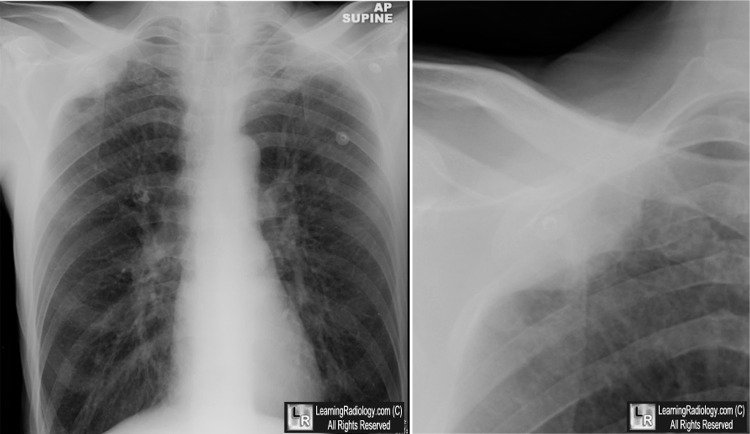
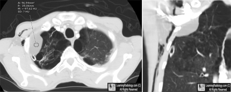
Pleural Lipoma. Above: White arrow points to a soft tissue mass in the right lung apex along the lateral chest wall which forms obtuse angles where it meets the chest wall (yellow arrows) suggesting a pleural or extrapleural location. Below: Axial CT shows the mass and demonstrates a density measurement equal to that of fat (red circle).
The coronal reconstruction gain shows the obtuse margins of the mass. The patient also had emphysema and a pneumothorax with subcutaneous emphysema.
For this same photo without the arrows, click here and here
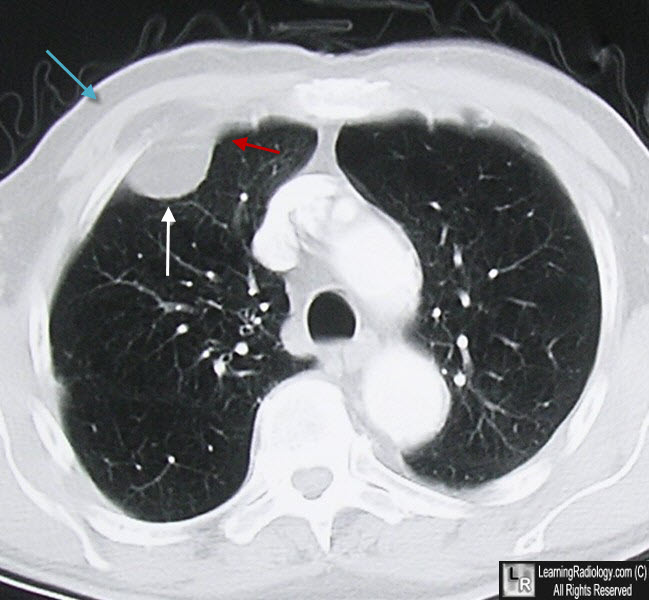
Pleural Lipoma. A mass (white arrow) of the same density as subcutaneous fat (blue arrow) produces an obtuse angle where it meets the chest wall (red arrow) suggesting it arises outside of the visceral pleura.
For more information, click on the link if you see this icon 
Fat-containing Lesions of the Chest. SC Gaerte, CA Meyer, HT Winer-Muram, RD Tarver, and DJ Conces Jr. October 2002, RadioGraphics, 22, S61-S78
|
|
|
